⏱️ Reading time: 7 min
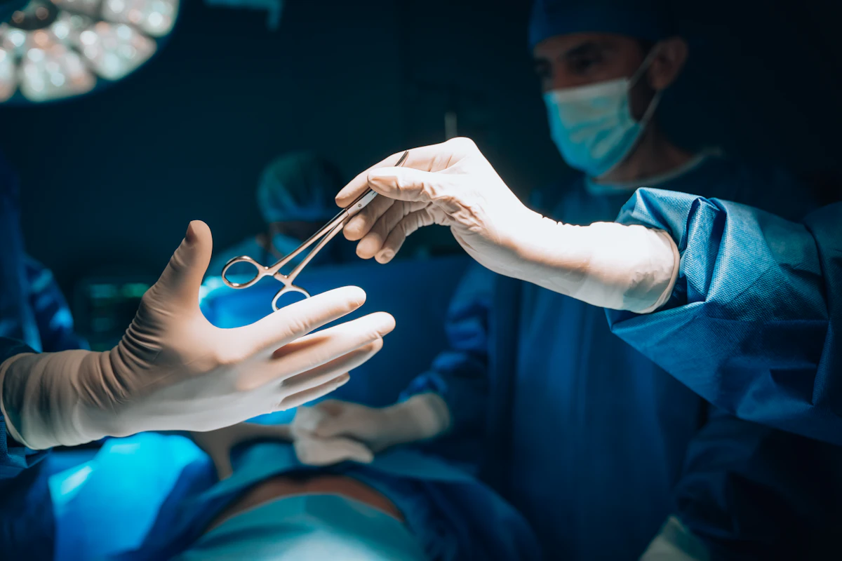
A surgeon watched a screen, right there in the operating room, showing exactly where a brain tumor ended and healthy tissue began, thanks to an artificial intelligence system that analyzed the images while he operated. That’s how People reported it in August 2026, calling the tool “Groundbreaking Technology”.
📑 En este artículo
The specific story has little public technical detail, but the technology category behind it is real and has been maturing for over a decade: AI-guided surgery built on neuronavigation and intraoperative imaging. This article explains exactly what problem this type of system solves and what open tools exist today for experimenting with brain tumor segmentation.
TL;DR
- People reported in August 2026 that surgeons used AI to guide the real-time removal of a brain tumor.
- The technology analyzes intraoperative images to mark the boundary between tumor and healthy tissue during the operation.
- Companies like Medtronic and Brainlab already sell neuronavigation systems that incorporate AI-assisted segmentation.
- Open frameworks like MONAI and nnU-Net let you train and run these segmentation models without relying on a closed vendor.
- The central technical challenge is “brain shift”: the brain deforms once the skull is opened, making pre-surgery images outdated.
- These systems are regulated as medical software (SaMD) and require clinical validation before being used on real patients.
- The open BraTS dataset is the community’s standard benchmark for evaluating brain tumor segmentation models.
What Happened
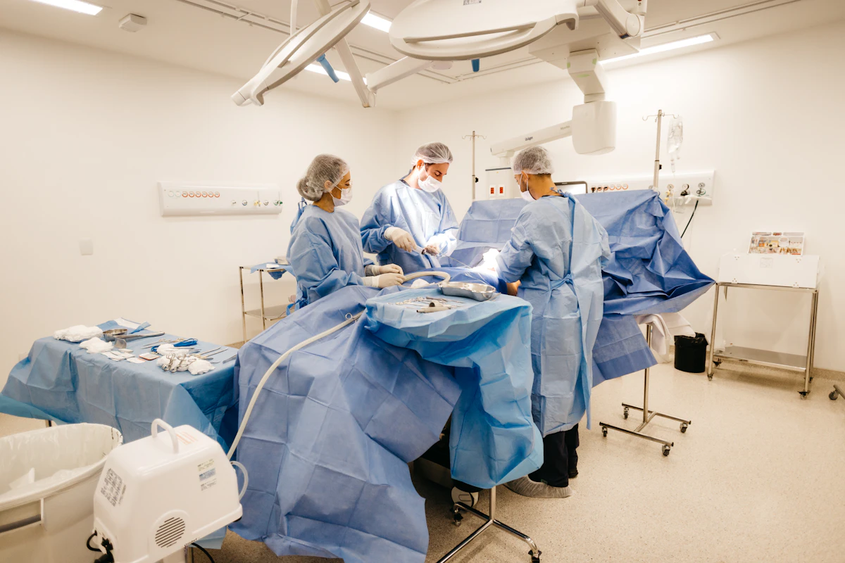
According to People’s coverage, a medical team used an artificial intelligence system to outline a brain tumor while the surgery was underway, instead of relying solely on the image taken before opening the skull. The original article doesn’t specify the hospital, the AI model, or the type of tumor, which is common in general-audience coverage of medical technology.
What can be explained precisely is the technology category that makes this kind of assistance possible: neuronavigation systems combined with automatic medical image segmentation. It’s not a new concept, but real-time AI segmentation is relatively recent in everyday clinical practice.
Context and History
Neuronavigation as a discipline was born in the 1990s: it involves registering an MRI scan taken before surgery and projecting it onto the real position of the surgical instruments, like a GPS for the brain. The persistent problem was that this prior image becomes outdated as soon as the surgeon opens the skull: cerebrospinal fluid drains, tissue sinks, and the brain shifts a few millimeters, a phenomenon the medical literature calls brain shift.
Intraoperative MRI (iMRI) emerged to correct that mismatch: an MRI scanner inside or near the operating room that rescans the patient midway through surgery. It’s precise, but expensive and slow: each new image can take minutes that the surgical team spends waiting. Automatic AI segmentation attacks that bottleneck, processing the new image and marking the tumor’s edge in seconds instead of having a radiologist draw it by hand.
Technical Details and Performance
How the Pipeline Works
The typical workflow for this type of system has four steps: acquiring the intraoperative image (MRI or ultrasound), preprocessing and normalization, running inference with a segmentation model trained to distinguish tumor tissue from healthy tissue, and overlaying the result on the surgeon’s view.
flowchart TD
A["Intraoperative image (MRI or ultrasound)"] --> B["Preprocessing and normalization"]
B --> C["Segmentation model (AI)"]
C --> D["Tumor tissue mask"]
D --> E["Overlay on surgeon's screen"]
E --> F["Live clinical decision"]
The two reference open source libraries in medical segmentation research are MONAI, a PyTorch-based framework maintained with NVIDIA and King’s College London, and nnU-Net, from the German Cancer Research Center (DKFZ), which automates network configuration based on the dataset. Neither one is, by itself, an approved clinical product: they are the foundation on which products like those from Medtronic or Brainlab are built.
💡 Tip: If you want to experiment with medical segmentation without access to real clinical data, the open BraTS (Brain Tumor Segmentation) dataset is the standard benchmark the community uses to train and evaluate these models.
Comparing Approaches
| Method | When It’s Used | Advantage | Limitation |
|---|---|---|---|
| Passive optical neuronavigation | Surgeries where tissue shifts little | Low cost, doesn’t require AI models | Loses accuracy with brain shift |
| Intraoperative MRI (iMRI) | Tumors with diffuse borders, like infiltrating gliomas | Updated imaging right inside the operating room | Requires an MRI scanner in the OR, high installation cost |
| Automatic AI segmentation | When the tumor’s edge needs to be marked in seconds | Reduces interpretation time during surgery | Requires clinical validation and doesn’t replace the surgeon’s judgment |
How to Start Trying It Out
To see the type of model behind these systems without needing real clinical data, you can install MONAI and run a minimal inference:
pip install monai nibabel

python -c "import monai; print(monai.__version__)"
That command installs the framework and confirms the active version. For a more realistic case, nnU-Net lets you run inference on an already preprocessed image volume:
nnUNetv2_predict \
  -i /data/patient_001/images \
  -o /data/patient_001/segmentation \
  -d 001 -c 3d_fullres -f 0
That command takes the images from the input folder and saves the resulting segmentation mask in the output folder. To confirm the model generated a real mask and not an empty file, you can read its dimensions directly:
python -c "import nibabel as nib; print(nib.load('segmentation/patient_001.nii.gz').shape)"If the result returns the same dimensions as the input image, the inference ran correctly across the entire volume.
⚠️ Heads up: No automatic segmentation system replaces the surgeon’s judgment or substitutes for the clinical validation regulators like the FDA require before use on real patients.
Impact and Analysis
The real value of AI-guided surgery isn’t replacing the surgeon, but compressing the time between “taking a new image” and “knowing where the tumor’s edge is”. That time, in a surgery lasting hours with the patient under anesthesia, carries a direct clinical cost.
The honest limit of this technology is validation: a segmentation model can fail in ways an experienced radiologist would catch but an automatic system won’t, especially in tumors with borders heavily infiltrated into healthy tissue. That’s why these systems are classified as medical software (SaMD) and go through regulatory processes before reaching the operating room; they aren’t deployed like just any app.
What’s Next
The visible trend in the industry is that open frameworks like MONAI and nnU-Net are closing the gap between academic research and clinical product: more and more papers publish weights and code alongside their results, which speeds up how quickly a lab finding reaches a product from a manufacturer like Medtronic or Brainlab, in fewer cycles than before.
Try it yourself: install MONAI with pip install monai and run the example dataset from its GitHub repository to see a medical segmentation pipeline running on your machine today.
Frequently Asked Questions
What exactly does the AI do during surgery?
It processes the intraoperative image and generates a mask marking which pixels or voxels correspond to tumor tissue, to overlay it on the screen the surgeon uses.
What is brain shift?
It’s the physical deformation the brain undergoes when the skull is opened and cerebrospinal fluid drains, which makes the MRI taken before surgery outdated.
What are MONAI and nnU-Net?
They are two open source frameworks for AI-driven medical imaging. MONAI is built on PyTorch, and nnU-Net automates the neural network configuration based on the dataset it’s given.
Are these systems already approved for general clinical use?
Commercial products from manufacturers like Medtronic or Brainlab go through regulatory processes as medical software (SaMD); open frameworks like MONAI and nnU-Net are research tools, not approved clinical products on their own.
Can someone without medical training experiment with this technology?
Yes, using public datasets like BraTS and frameworks like MONAI or nnU-Net, it’s possible to train and run brain tumor segmentation models without access to real clinical data.
References
- People / MSN: original report on the use of AI to guide the real-time removal of a brain tumor.
- MONAI: open source PyTorch-based framework for AI-driven medical imaging.
- nnU-Net (GitHub): biomedical segmentation framework from the German Cancer Research Center.
- Wikipedia: Neuronavigation: history and fundamentals of image-guided surgery.
- Medtronic: manufacturer of neuronavigation systems used in image-guided surgery.
📱 Like this content? Follow @programacion on Telegram for daily tech content in Spanish: quick summaries, fresh content every day.
Imagen destacada: Foto de Navy Medicine en Unsplash
Did it work for you? Got a different error? Say so below: questions get answered and help the next reader.
Leave a comment


0 Comments